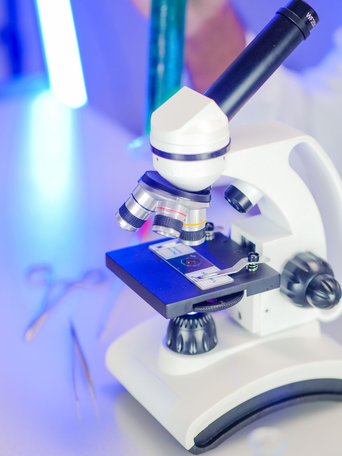
Total internal reflection fluorescence microscopy (TIRFM) uses the evanescent wave from total internal reflection to selectively excite fluorophores within 100-200 nanometres of the coverslip. This eliminates background fluorescence from deeper layers and produces images with extraordinary signal-to-noise. It is the method of choice for watching vesicles fuse with the cell membrane, tracking single molecules, and studying events at the surface of living cells.
Picture a scientist trying to watch a single protein docking at a cell membrane using a standard fluorescence microscope. The whole cell glows, drowning the surface signal in haze from the interior. TIRF solves this by illuminating only the bottom 100 nm of the cell — roughly one-thousandth the thickness of a human hair. Everything above stays dark, and the surface event becomes crystal clear.
What is TIRF microscopy?
TIRF microscopy is a fluorescence imaging technique that exploits total internal reflection to create an extremely thin excitation zone at the interface between a glass coverslip and the aqueous sample. The technique was first described by Hirschfeld in 1965 and developed into a practical biological tool by Daniel Axelrod in the 1980s.
The core idea is simple. In conventional epifluorescence microscopy, the entire sample is bathed in excitation light, producing bright fluorescence from everywhere — including blurry out-of-focus contributions that degrade the image. TIRF limits excitation to within about 100 nm of the coverslip by using an evanescent wave instead of a propagating light beam. Only fluorophores within that evanescent zone are excited. Everything else remains dark.
This selective illumination makes TIRF ideal for studying processes at or near the cell membrane, including endocytosis, exocytosis, cell adhesion, cytoskeletal dynamics, and receptor-ligand interactions. It is also a cornerstone technique in single-molecule biophysics.
The Wikipedia entry on TIRF microscopy provides a thorough overview of the history and technical details.
How TIRF works: the evanescent wave
TIRF relies on the same physics as fibre optics and diamond sparkle: total internal reflection. When light travelling through glass hits the interface with water or cell culture medium at an angle greater than the critical angle, it reflects entirely back into the glass. But a faint electromagnetic field — the evanescent wave — extends into the aqueous medium.
The evanescent wave has two properties that make TIRF possible:
It decays exponentially. The intensity of the evanescent wave drops off with distance from the interface according to an exponential decay. The characteristic penetration depth — the distance at which the intensity falls to 1/e of its surface value — is typically 100-200 nm. This depends on the wavelength of the light, the angle of incidence, and the refractive indices of the glass and the sample medium.
It excites fluorophores identically to a normal light beam. Despite being non-propagating in the direction away from the interface, the evanescent wave oscillates at the same frequency as the incident laser light. It can excite fluorescent molecules just as a transmitted beam would. The fluorescence emitted by excited molecules is then collected by the microscope objective and forms the TIRF image.
The critical angle that makes this possible depends on the refractive indices of the two media. For a glass-water interface, the critical angle is about 61 degrees — different from the 48.6 degrees for water to air because the surrounding medium is water (n = 1.33), not air.
Our guide on total internal reflection explains the conditions for TIR and the critical angle formula in detail. The critical angle calculator and worked examples show how to compute the threshold for different material pairs.
Two TIRF configurations
There are two common ways to generate the evanescent wave for TIRF microscopy:
Prism-based TIRF
The original configuration uses a prism to direct the laser beam toward the coverslip interface at the required angle. The prism is optically coupled to the coverslip above the sample. This method is simple and inexpensive, and it was the standard approach for the first 30 years of TIRF use.
The disadvantage: the prism sits above the sample, blocking access. You cannot manipulate the specimen, inject reagents, or use electrophysiology tools while imaging. This made prism-based TIRF impractical for many live-cell experiments.
Objective-based TIRF
Modern TIRF systems use a high-numerical-aperture objective (typically NA ≥ 1.45) to direct the laser light at the correct angle through the objective itself. The laser beam enters the objective off-centre, exits at an oblique angle, and strikes the coverslip-sample interface beyond the critical angle.
Objective-based TIRF leaves the space above the sample completely free for micromanipulators, perfusion systems, and other tools. It is compatible with standard inverted microscopes and has become the dominant configuration for biological TIRF imaging.
The Nikon MicroscopyU guide on TIRF provides detailed diagrams comparing both configurations and explains how the evanescent wave intensity profile changes with the incidence angle.
Advantages of TIRF microscopy
TIRF offers several advantages over conventional fluorescence microscopy techniques:
Low background. Only the thin evanescent zone is illuminated, so there is almost no out-of-focus fluorescence. Signal-to-noise ratios are dramatically higher than in epifluorescence.
Low photobleaching and phototoxicity. Because most of the cell is not exposed to excitation light, fluorophores last longer and cells stay healthier during long timelapse experiments.
Fast acquisition. TIRF is a widefield technique — the entire field of view is imaged simultaneously onto a camera sensor. This allows frame rates far faster than confocal or multiphoton microscopy, which scan point by point.
Single-molecule sensitivity. The combination of low background and efficient excitation makes TIRF one of the best techniques for visualising individual fluorescent molecules. It is widely used in single-molecule tracking, super-resolution microscopy, and studies of molecular kinetics.
The main limitation is that TIRF only images structures within about 200 nm of the coverslip. It cannot image deep inside cells or through tissue slices. For deeper imaging, confocal or multiphoton microscopy are better choices.
Applications of TIRF microscopy
TIRF has become an essential tool in cell biology and biophysics. Common applications include:
Vesicle trafficking. TIRF excels at watching individual vesicles approach, dock, and fuse with the plasma membrane during exocytosis and endocytosis. The thin excitation zone means only vesicles near the membrane are visible, and the fusion event itself can be tracked in real time.
Cell adhesion. Focal adhesions — the complexes that anchor cells to the substrate — lie within the evanescent zone. TIRF reveals their assembly, disassembly, and dynamics with high contrast.
Cytoskeletal dynamics. Actin filaments and microtubules near the cell surface can be imaged with TIRF, revealing the polymerization and depolymerisation events that drive cell shape changes and movement.
Single-molecule imaging. The low background of TIRF makes it possible to detect and track individual fluorescent molecules. This has been used to study receptor diffusion in the membrane, motor protein stepping, and enzyme kinetics at the single-molecule level.
The News-Medical guide on TIRF microscopy discusses several of these applications with example images and practical protocols.
TIRF vs other microscopy techniques
TIRF vs epifluorescence: Epifluorescence illuminates the entire sample depth. TIRF restricts excitation to 100-200 nm. TIRF wins for surface imaging; epifluorescence is needed for deeper structures.
TIRF vs confocal: Confocal uses a pinhole to reject out-of-focus light and can image at any depth, but it scans point by point (slower) and exposes the entire sample to excitation light (more photobleaching). TIRF is faster, gentler, and provides better axial confinement at the surface, but cannot image deeper than ~200 nm.
TIRF vs FRAP: Fluorescence recovery after photobleaching (FRAP) measures molecular mobility. TIRF can be combined with FRAP to measure diffusion specifically at the membrane, where conventional FRAP would also bleach the interior.
Common misconception: TIRF requires a specialised microscope
Many people assume TIRF requires purpose-built, expensive hardware. Objective-based TIRF can be achieved on a standard inverted microscope by adding a TIRF illuminator module and a high-NA objective. Many modern research microscopes from Nikon, Olympus, Zeiss, and Leica offer TIRF as an upgrade option. That said, a complete TIRF system with laser launch, illuminator, high-NA objective, and sensitive camera still represents a significant investment — typically $100,000-$300,000 depending on the configuration.
For a detailed look at how reflection and refraction underpin TIRF and other optical techniques, see our comparison of reflection vs refraction.
Frequently Asked Questions
What is TIRF microscopy?
TIRF (total internal reflection fluorescence) microscopy uses an evanescent wave generated by total internal reflection at the glass-sample interface to selectively excite fluorophores within 100-200 nm of the coverslip. This produces high-contrast images of cell membrane and near-surface processes with minimal background fluorescence.
What is the difference between TIRF and confocal microscopy?
TIRF uses an evanescent wave to illuminate only a 100-200 nm slice adjacent to the coverslip, while confocal uses pinholes to reject out-of-focus light from deeper in the sample. TIRF provides better axial resolution near the surface and much lower photobleaching, but confocal can image deeper into the sample.
How does TIRF microscopy work?
In TIRF microscopy, laser light is directed at the glass-sample interface at an angle exceeding the critical angle. Total internal reflection occurs, generating an evanescent wave that penetrates only 100-200 nm into the sample. This evanescent wave selectively excites fluorophores in this thin region, producing fluorescence with almost no background from deeper layers.
What are the advantages of TIRF microscopy?
TIRF provides outstanding signal-to-noise ratio by eliminating out-of-focus background fluorescence. It causes much less photobleaching and phototoxicity than widefield or confocal because only a thin region is illuminated. It is also a widefield technique, allowing rapid image acquisition suitable for live-cell imaging.
What is the evanescent wave in TIRF?
The evanescent wave is an electromagnetic field generated at the interface during total internal reflection. Its intensity decays exponentially with distance from the interface. In TIRF microscopy, the penetration depth is typically 100-200 nm, meaning only fluorophores within this region are efficiently excited.
What is the difference between prism-based and objective-based TIRF?
Prism-based TIRF directs the laser through a prism above the sample, which limits access to the specimen for manipulations. Objective-based TIRF uses a high-NA objective to achieve TIR through the objective itself, providing easier access to the sample and compatibility with standard inverted microscopes. Objective-based is now the more common configuration.

About Umar Farooq
Umar Farooq writes in-depth guides on the physics of light and optics — from reflection, refraction, and lenses to diffraction, lasers, and fiber optics, explained from first principles.
Read more about Umar Farooq →