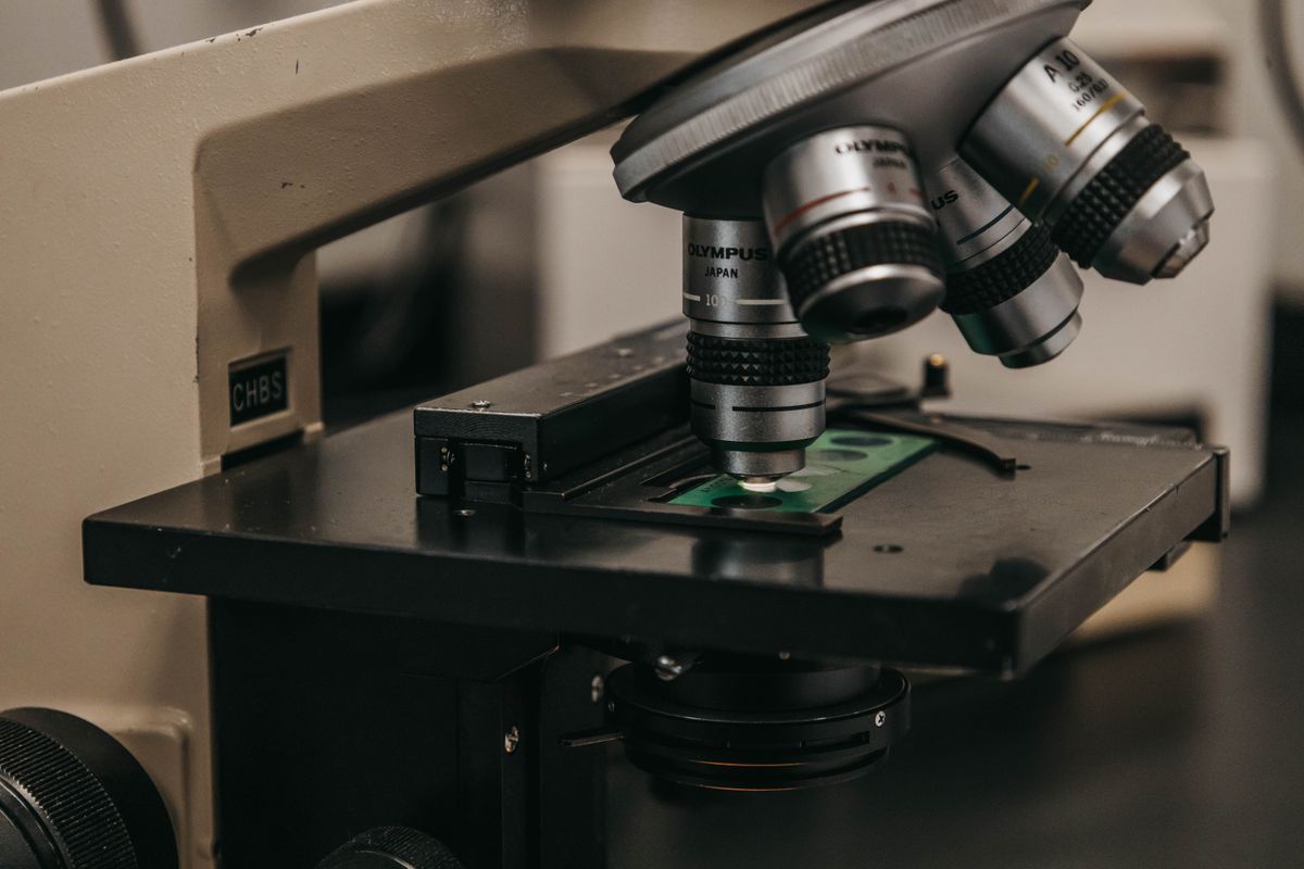
A diffraction grating spectrometer is the instrument that turns light into data. Point it at a flame and it tells you which elements are burning. Point it at a star and it reveals what the star is made of. Point it at a gemstone and it distinguishes a natural diamond from a laboratory-grown one. The instrument does all of this by one core trick: it separates light into its constituent wavelengths and measures their intensities.
Think of white light as a crowd leaving a stadium — all the colours are mixed together. The spectrometer is the turnstile system that directs each colour into its own lane, then counts how many people went through each lane. The grating does the sorting. The detector does the counting.
How a diffraction grating spectrometer works
The optical path inside a spectrometer follows five steps:
- Entrance slit. Light from the source passes through a narrow slit. The slit width controls the trade-off between resolution and brightness — a narrower slit gives sharper spectral lines but less light reaches the detector.
- Collimator. A lens or concave mirror converts the diverging light from the slit into a parallel beam. This ensures that all rays strike the grating at the same angle.
- Diffraction grating. The parallel beam hits the grating, which splits it into different wavelengths travelling at different angles. The angle for each wavelength follows the grating equation: d sin θ = mλ, where d is the groove spacing, θ is the diffraction angle, m is the order, and λ is the wavelength.
- Focusing mirror. A second mirror or lens collects the diffracted beams and focuses them onto the detector.
- Detector. A CCD or CMOS sensor records the intensity of light at each position. Each pixel corresponds to a narrow range of wavelengths. The result is a spectrum: a graph of intensity versus wavelength.
In a simple spectroscope (the visual kind), step 5 is replaced by an eyepiece that lets you see the spectrum directly. No numbers, no graph — just the coloured lines.
Spectrometer vs spectroscope vs monochromator
These three terms are often confused. They share the same basic optical layout but differ in what they output:
- Spectroscope: visual only. You look through an eyepiece and see the spectrum with your eye. Common in classrooms and gemology.
- Spectrometer: quantitative. A detector records intensity as a function of wavelength, producing digital data you can analyse.
- Monochromator: has an exit slit that selects a single narrow wavelength band. Used when you need to illuminate a sample with one colour at a time — for example, in fluorescence measurements or laser tuning. The grating rotates to scan different wavelengths across the exit slit.
A monochromator is essentially a spectrometer used in reverse: instead of measuring all wavelengths at once, it outputs one wavelength at a time.
Types of spectrometer designs
Czerny-Turner
The most common laboratory design. Light goes from the entrance slit to a collimating mirror, then to a planar diffraction grating, then to a focusing mirror, then to the detector. The two mirrors eliminate chromatic aberration (which lenses would introduce). Most research-grade UV-Vis spectrometers use the Czerny-Turner layout.
Echelle
An Echelle spectrometer uses a coarse grating (low groove density) at a very high diffraction order, combined with a second dispersive element (a prism or another grating) to separate the overlapping orders. This gives very high resolution in a compact package — ideal for applications like astrophysics where you need to resolve closely spaced spectral lines.
Concave grating
A single concave grating serves as both the dispersive element and the focusing mirror, eliminating the need for separate collimating and focusing optics. This simplifies the design and reduces light loss. Common in compact fibre-optic spectrometers used for field measurements.
How spectral resolution works
The resolution of a grating spectrometer — its ability to distinguish two nearby wavelengths — depends on three factors:
- Groove density (lines per millimetre). More lines per mm spreads the spectrum wider, improving resolution.
- Illuminated width. The wider the beam on the grating, the more grooves participate in the diffraction. The total number of illuminated grooves N = (beam width) × (groove density).
- Diffraction order. Higher orders give proportionally higher resolution (though with lower intensity).
The fundamental resolution is: R = λ / Δλ = mN
A typical laboratory spectrometer might have a 50 mm wide grating with 600 lines/mm, giving N = 30,000 grooves. In first order (m = 1), R = 30,000. This means it can resolve two spectral lines separated by 0.02 nm at 600 nm — more than enough to separate the sodium D doublet at 589.0 nm and 589.6 nm.
In practice, the resolution is also limited by the entrance slit width. A slit that is too wide reduces resolution even if the grating is capable of more. This is why high-resolution spectrometers have very narrow slits (typically 10–50 µm).
Calibration: how you know the wavelength
A spectrometer does not directly measure wavelength. It measures the position of a spectral line on the detector. To convert pixel position to wavelength, you calibrate using a source with known spectral lines.
A mercury vapour lamp is the standard calibration source. It emits bright, well-characterised lines at 404.7 nm (violet), 435.8 nm (blue), 546.1 nm (green), and 577.0/579.1 nm (yellow). By measuring which pixels these lines fall on, you create a calibration curve that maps every pixel to a wavelength. Sodium lamps (589.0/589.6 nm doublet) and neon lamps are also common.
Without calibration, the wavelength values from a spectrometer are only approximate. With calibration, they are accurate to a fraction of a nanometre.
5 uses of diffraction grating spectrometers
1. Chemistry: identifying elements. Every element emits a unique set of spectral lines. A spectrometer identifies which elements are present in a sample by matching its emission lines to a reference database. Used in materials analysis, forensics, and environmental testing.
2. Astronomy: analysing starlight. A spectrograph attached to a telescope splits starlight into its spectrum. The absorption lines reveal the star's chemical composition, temperature, density, and radial velocity (via Doppler shift). This is how we know what the Sun and distant stars are made of.
3. Gemology: identifying gemstones. A hand-held spectroscope lets gemologists distinguish natural gems from synthetics by their absorption spectra. For example, natural emerald shows a distinct chromium absorption line that synthetic emerald lacks. The GIA (Gemological Institute of America) sells a diffraction grating spectroscope specifically for this purpose.
4. Microscopy: microspectroscopy. Coupling a spectrometer to a microscope lets you measure the spectrum of a microscopic area — a single cell, a pigment grain in a painting, or a mineral inclusion in a rock section. This is used in biomedical imaging, art conservation, and geology.
5. Environmental monitoring. Spectrometers measure pollutants in air and water by detecting their characteristic absorption bands. NO₂, ozone, and sulphur dioxide all have distinct spectral signatures in the UV and visible range.
For more on the physics of diffraction itself, start with our guide on what is diffraction. The what is a diffraction grating article covers the grating component itself in detail. To understand the Fraunhofer diffraction regime that makes spectrometer optics possible, see Fraunhofer vs Fresnel diffraction.
External resources
- Ossila: How Do Spectrometers Work? — clear guide covering entrance slit, grating, and detector with practical advice on choosing a spectrometer
- Shimadzu: Introduction to Diffraction Gratings — technical resource from a major spectrometer manufacturer
- Physics Forums: Fundamentals of the Diffraction Grating Spectrometer — deep dive into the physics and resolution of grating spectrometers
Frequently Asked Questions
What is a diffraction grating spectrometer?
A diffraction grating spectrometer is an optical instrument that separates light into its component wavelengths using a diffraction grating. It consists of an entrance slit, a collimator, a diffraction grating, and a detector (or eyepiece). It is used to measure the wavelengths and intensities of spectral lines emitted or absorbed by substances, allowing identification of chemical elements and molecular structures.
How does a diffraction grating spectrometer work?
Light enters through a narrow slit and is collimated into a parallel beam by a lens or mirror. The parallel beam strikes a diffraction grating, which splits it into different wavelengths at different angles based on the grating equation d sin θ = mλ. The separated wavelengths are then focused onto a detector — either a CCD array for digital measurement or an eyepiece for visual observation. By measuring the angles of the diffracted beams, the wavelengths of the spectral lines can be calculated.
What is the difference between a spectrometer, spectroscope, and monochromator?
A spectroscope is a simple visual instrument that lets you see a spectrum through an eyepiece. A spectrometer is a quantitative instrument that measures wavelengths and intensities using a detector. A monochromator is a type of spectrometer with an exit slit that isolates a single narrow wavelength band, used when you need to select one colour at a time for an experiment.
What is a diffraction grating under a microscope?
A diffraction grating under a microscope refers to the technique of placing a grating in the optical path of a microscope, often in the form of a grating spectrometer coupled to a microscope. This setup, called microspectroscopy, measures the absorption or emission spectrum of microscopic samples — such as a single cell, a grain of pigment in a painting, or a microscopic crystal in a mineral section.
What is the resolution of a diffraction grating spectrometer?
The spectral resolution of a grating spectrometer is given by R = λ / Δλ = mN, where m is the diffraction order and N is the total number of illuminated grooves. A 50 mm wide grating with 600 lines/mm in first order gives R = 50 × 600 = 30,000, meaning it can resolve two spectral lines separated by 0.02 nm at 600 nm. Higher resolution requires a wider grating, higher groove density, or a higher diffraction order.
What are the uses of a diffraction grating spectrometer?
Key uses include: identifying chemical elements in materials science, analysing starlight in astronomy, gemstone identification in gemology, monitoring pollutants in environmental science, measuring oxygen saturation in medical pulse oximetry, and quality control in pharmaceutical manufacturing. Anywhere you need to know what wavelengths of light a substance emits or absorbs, a spectrometer is the tool.

About Umar Farooq
Umar Farooq writes in-depth guides on the physics of light and optics — from reflection, refraction, and lenses to diffraction, lasers, and fiber optics, explained from first principles.
Read more about Umar Farooq →