
How does a confocal microscope work? It works by focusing a laser to a single point in the sample, using a pinhole to block any light coming from above or below that point, and scanning across the sample to build a full image one pixel at a time. The pinhole is the key. It sits at a plane that is optically conjugate to the focal plane — hence "confocal" — and it rejects everything except light that originates exactly where the laser is focused. This gives the confocal microscope a superpower: it can take sharp, thin optical sections through thick specimens without ever cutting them.
Here is how it works, from the laser to the final image.
What is a confocal microscope?
A confocal microscope is a type of fluorescence microscope that uses point illumination and a pinhole spatial filter to eliminate out-of-focus light. In a conventional widefield fluorescence microscope, the entire sample is bathed in excitation light. Everything fluoresces at once — the in-focus parts and the out-of-focus parts — so the image has a hazy background that obscures fine detail, especially in thick specimens.
Here is a way to picture the difference. Imagine trying to hear one conversation in a crowded room. A widefield microscope is like listening to the whole room — you hear the conversation you want, but also every other conversation, the clatter of glasses, and the footsteps. A confocal microscope is like putting a cardboard tube to your ear and aiming it at just one person. You hear that person clearly and almost nothing else.
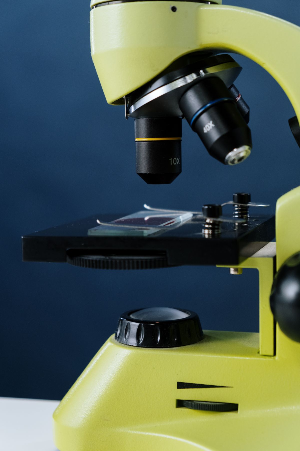
The tube is the pinhole. And the person is the single point where the laser is focused.
The key components
A confocal laser scanning microscope has five essential parts:
- A laser — provides intense, single-wavelength excitation light that can be focused to a tiny spot.
- Scanning mirrors — two galvanometer-driven mirrors that steer the laser beam in a raster pattern across the sample.
- A dichroic mirror — reflects the excitation beam toward the sample but transmits the longer-wavelength fluorescence coming back.
- A pinhole aperture — sits in front of the detector at a plane conjugate to the focal plane. This is the heart of the confocal design.
- A photomultiplier tube (PMT) — a highly sensitive detector that converts photons into an electrical signal.
The arrangement is elegant. The laser reflects off the dichroic, through the scanning mirrors and objective, and into the sample. The fluorescence comes back along the same path, passes through the scanning mirrors (this "descans" the beam — keeps it stationary at the pinhole), through the dichroic, and is focused onto the pinhole. The PMT sits behind the pinhole and measures whatever light gets through.
How the pinhole creates optical sections
The pinhole is what makes a confocal microscope "confocal." The name comes from the fact that the pinhole lies in a plane that is conjugate to the focal plane of the objective. Any light that originates at the focal point in the sample is focused to a small spot right at the pinhole, so it passes through to the detector.
Light that originates from above or below the focal plane? It is also focused — but to a spot above or below the pinhole plane. That spot is larger and defocused at the pinhole position, so most of it hits the edges and is blocked. Only a tiny fraction slips through.
The effect is dramatic. A confocal microscope rejects out-of-focus fluorescence from regions as little as 0.5 micrometres above or below the focal plane. This is what makes optical sectioning possible. Instead of physically slicing a specimen into thin sections — which kills cells and takes hours — the microscope can capture a sharp image from a single plane within a living, intact sample. By moving the stage up in small steps and repeating, you collect a stack of optical sections called a z-stack.
The how does a lens work article explains the basics of how objective lenses focus light, which is the foundation of what a confocal objective does.
Scanning: building the image point by point
At any given instant, a laser scanning confocal microscope is only observing a single point in the sample — about 0.25 to 0.8 micrometres in diameter. To build a complete image, the beam must scan across the sample.
Two galvanometer mirrors do this. One steers the beam left and right (the x-axis). The other steers it up and down (the y-axis). Together they trace a raster pattern — the same pattern an old CRT television used — across the sample. A typical 512 × 512 pixel image takes about one second to acquire at standard scan speeds.
As the beam moves, the fluorescence intensity at each point varies. The PMT converts these variations into an electrical signal. An analog-to-digital converter turns that signal into pixels. The computer assembles the pixels into an image. Because only one point is observed at a time, the image never exists as a real optical image — it is reconstructed by the computer from the stream of measurements.
This point-scanning design has a useful side effect: built-in zoom. By scanning a smaller area, the microscope effectively magnifies the image electronically, without changing objectives.
Fluorescence: making the invisible visible
Most confocal microscopy relies on fluorescence. The specimen is stained with fluorescent dyes called fluorophores that absorb light at one wavelength and emit it at a longer one. The laser provides the excitation. The dichroic mirror separates the emitted fluorescence from the excitation beam.
The key advantage is that fluorescence from outside the focal plane is rejected by the pinhole — so images of thick specimens are crisp instead of blurry. This has made confocal microscopy invaluable for cell biology, where researchers need to see fine structures within living cells and tissues.
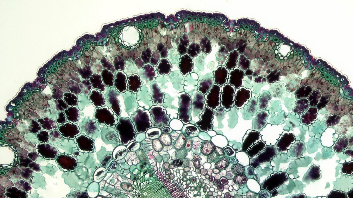
Common applications include imaging the cytoskeleton, tracking fluorescently tagged proteins in live cells, mapping neuronal networks, and studying biofilms. The technique is also used in materials science for surface profiling and in ophthalmology for corneal imaging. How a microscope works covers the basics of standard optical microscopy for comparison.
Types of confocal microscopes
Not all confocal microscopes are point scanners. There are two main types:
Laser scanning confocal microscopes (LSCM) — the kind described above. They use a single laser beam scanned by mirrors. They offer the highest resolution and flexible zoom, but scan speeds are limited to about one frame per second at full resolution.
Spinning-disk confocal microscopes — these use a Nipkow disk: a spinning wheel with hundreds of pinholes arranged in a spiral pattern. Multiple points are illuminated and detected simultaneously, so the image forms in real time. This reduces phototoxicity significantly, making spinning-disk systems the preferred choice for live-cell imaging. The trade-off is lower peak resolution and a fixed sampling density set by the camera.
Microlens-enhanced spinning-disk systems add a second disk of microlenses that focus light into each pinhole, boosting the signal and making them even better for low-light applications.
A quick clarification
People sometimes assume that a confocal microscope achieves better resolution than a scanning electron microscope (SEM). It does not. TEM and SEM resolve details down to a few nanometres; confocal microscopy is still diffraction-limited at roughly 200–300 nanometres laterally. The real advantage of confocal is not raw resolution — it is the ability to see deep inside living, intact specimens with fluorescence contrast, something no electron microscope can do. The two techniques are complementary, not competitors.
The how does an electron microscope work article covers SEM and TEM in detail and is worth reading alongside this one for a full picture.
History in brief
The confocal principle was patented by Marvin Minsky in 1957 — yes, the same Minsky who later co-founded the MIT AI Lab. He wanted to image neural networks in thick brain tissue but had no laser (the laser was invented three years later) and no computer fast enough to make it practical. His invention sat mostly unnoticed for two decades.
The first practical laser scanning confocal microscope was built by M. David Egger and Paul Davidovits in 1969. The technique took off in the 1980s as affordable lasers, computers, and sensitive detectors became available. The first commercial instruments appeared in 1987. Today, confocal microscopy is a standard tool in thousands of biology labs worldwide.
External resources
- Nikon's MicroscopyU — Introductory Confocal Concepts (verified, a clear primer on how confocal imaging works)
- Wikipedia — Confocal microscopy (verified, comprehensive technical reference)
Frequently Asked Questions
How does a confocal microscope work step by step?
A confocal microscope works in six steps: (1) A laser beam is focused to a diffraction-limited spot on the sample. (2) The sample fluoresces, emitting light at a longer wavelength. (3) That fluorescence passes back through the objective and is descanned by the same mirrors. (4) The light reaches a dichroic mirror that separates it from the excitation beam. (5) It passes through a pinhole aperture that blocks any light originating from above or below the focal plane. (6) A photomultiplier tube detects the filtered light, and a computer builds the image one pixel at a time as the beam scans across the sample.
What is the difference between a confocal microscope and a normal microscope?
A normal (widefield) microscope floods the entire sample with light, so fluorescence from above and below the focal plane creates a hazy background. A confocal microscope uses a focused laser spot and a pinhole to block out-of-focus light, producing much sharper images from a single optical plane. This allows optical sectioning — capturing thin slices through a thick specimen without physically cutting it.
What is the principle of confocal microscopy?
The principle is point illumination and point detection. A laser excites a single point in the sample. The emitted fluorescence passes through a pinhole placed at a conjugate focal plane, which blocks light from out-of-focus regions. Only in-focus light reaches the detector. The beam scans across the sample point by point to build the full image.
What is the pinhole in a confocal microscope?
The pinhole is a small circular aperture placed in front of the detector, at a plane conjugate to the focal plane of the objective. It acts as a spatial filter: light from the focal plane is focused through the pinhole, while light from above or below the focal plane forms a larger, defocused spot that is mostly blocked. Adjusting the pinhole diameter controls the thickness of the optical section.
Do confocal microscopes use lasers?
Most confocal microscopes use lasers as their excitation source. Lasers provide intense, monochromatic, coherent light that can be focused to a diffraction-limited spot — essential for point-scanning confocal microscopy. Common laser lines include argon-ion (488 nm, blue-green), helium-neon (543 nm, green; 633 nm, red), and diode lasers (405 nm, violet).
What is optical sectioning in confocal microscopy?
Optical sectioning is the ability to capture sharp images from a single thin plane within a thick specimen, without physically slicing it. By blocking out-of-focus light with the pinhole, a confocal microscope restricts the detected signal to a plane typically 0.5–1.5 micrometres thick. By moving the focal plane along the z-axis and collecting a series of these sections, the microscope can build a three-dimensional reconstruction of the sample.
Can a confocal microscope see live cells?
Yes. Confocal microscopy is widely used for live-cell imaging because it does not require physical sectioning. However, laser intensity must be kept low to avoid phototoxicity and photobleaching. Spinning-disk confocal microscopes are often preferred for live-cell work because they distribute the excitation across many points simultaneously, reducing the energy per point.
What are the advantages of confocal microscopy?
The main advantages are: (1) rejection of out-of-focus light for sharper images, (2) optical sectioning through thick specimens, (3) ability to reconstruct 3D volumes from z-stacks, (4) improved resolution — especially along the axial direction — compared to widefield fluorescence, and (5) the ability to image living specimens non-invasively.

About Umar Farooq
Umar Farooq writes in-depth guides on the physics of light and optics — from reflection, refraction, and lenses to diffraction, lasers, and fiber optics, explained from first principles.
Read more about Umar Farooq →