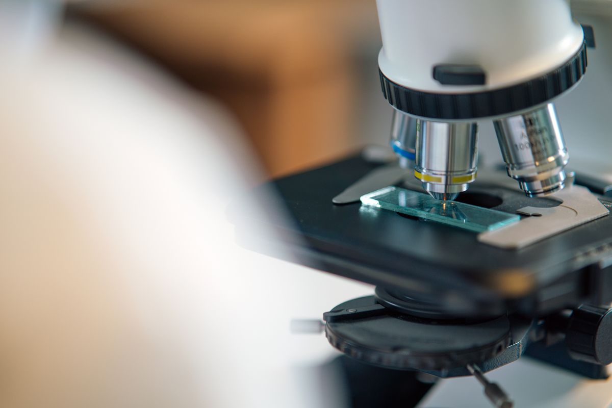
If you are wondering how does a light microscope work, the answer is straightforward. A light microscope passes visible light through a thin specimen and uses two glass lenses — an objective and an eyepiece — to magnify the image. The objective creates a magnified real image inside the tube, and the eyepiece magnifies that image for your eye. The total magnification is simply the objective power multiplied by the eyepiece power: a 10× eyepiece with a 40× objective gives 400×.
Here is what we cover: the optical path from light source to eye, how magnification and resolution differ, the main types of optical microscopes, and how they compare with electron microscopes.
What Is a Light Microscope?
A light microscope (or optical microscope) uses visible light and glass lenses to form an image. It is the oldest type of microscope, dating to the 17th century, and remains the most widely used in biology and medicine.
Light microscopes magnify up to about 1000-1500× and resolve details down to about 0.2 micrometres — roughly 200 nanometres, or about 500 times thinner than a human hair. The defining feature is that they use photons rather than electrons, which means they can image living cells in their natural state. No electron microscope can do that.
How Does a Light Microscope Work? The Optical Path
Picture a magnifying glass and a projector working as a team. The objective lens acts like a tiny projector, creating a large image of the specimen inside the microscope. The eyepiece then acts as a magnifying glass, letting you inspect that projected image up close.
Here is the step-by-step path light takes through a light microscope:
- Light source — An LED or halogen bulb shines light upward through the condenser.
- Condenser — A lens system focuses the light into a cone that illuminates the specimen evenly.
- Specimen — Light passes through the thin specimen on the glass slide.
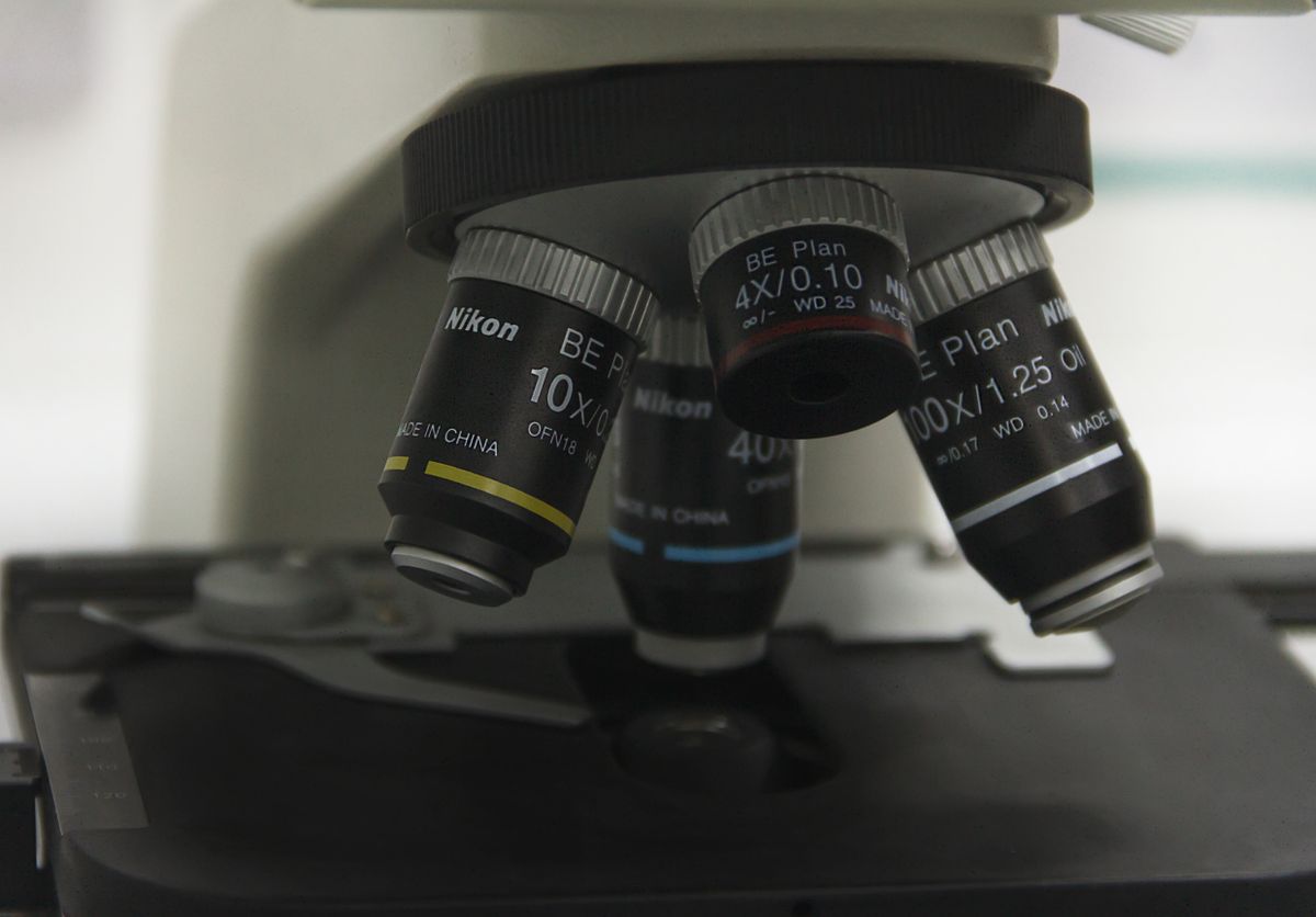
- Objective lens — This is the primary magnifying lens. It collects the transmitted light and forms a real, inverted, enlarged image inside the microscope tube. Objectives are marked with their magnification (4×, 10×, 40×, 100×) and numerical aperture (NA), which determines resolution.
- Eyepiece — This acts as a magnifying glass, enlarging the intermediate image further (typically 10×). The final image your eye sees is virtual — it appears to be behind the lens.
- Your eye — The lens in your eye focuses the virtual image onto your retina.
The two-stage design is the key to how does a light microscope work. A single lens cannot reach high magnifications without severe distortion. Splitting the work between objective and eyepiece lets each lens do what it does best.
Magnification in a Light Microscope
Total magnification follows a simple rule:
Total magnification = objective magnification × eyepiece magnification
A standard 10× eyepiece with a 40× objective gives 400× total magnification. With a 100× oil-immersion objective, you reach 1000×.
The eyepiece is almost always 10×, so your real choice is the objective. Low-power objectives (4×, 10×) give a wide field of view for finding your specimen. High-power objectives (40×, 100×) show finer detail but need careful focusing and good lighting.
Oil-immersion objectives use a drop of oil between the lens and the slide. The oil has the same refractive index as crown glass (about 1.52), which prevents light from bending as it leaves the slide. This lets the objective collect more light and achieve higher resolution.
Resolution: The Real Limit
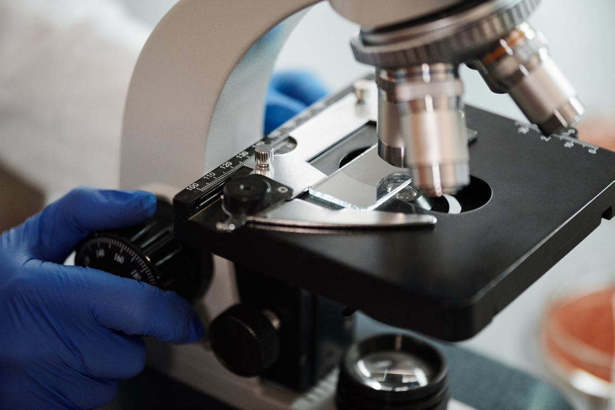
A common mistake is to think magnification is the point of a microscope. It is not. Resolution — the ability to distinguish two close points as separate — matters far more. Magnifying a blur just gives you a bigger blur.
In 1873, the German physicist Ernst Abbe discovered that no optical microscope can resolve objects smaller than roughly half the wavelength of the light it uses. This is the Abbe diffraction limit:
Resolution limit ≈ λ / (2 × NA)
For green light (λ ≈ 500 nm) and a good objective (NA ≈ 1.4), the resolution limit is about 0.18 µm — roughly 180 nm. This is a hard physical limit. Details smaller than that are invisible no matter how much you magnify.
The practical takeaway: a light microscope at 1000× magnification is enough to see bacteria (1-5 µm), cell nuclei (about 10 µm), and mitochondria (0.5-1 µm). Going beyond 1000× on a standard instrument gives empty magnification — the image gets bigger but no new detail appears, like zooming into a low-resolution photo.
This is why electron microscopes exist. By using electrons with wavelengths about 100,000 times shorter than visible light, they achieve resolution down to 0.1 nm — roughly 2000 times better than any light microscope.
Types of Light Microscopes
Not all light microscopes work the same way. Different techniques reveal different information:
- Brightfield — The standard type described above. Specimens appear dark against a bright background. Best for stained or naturally coloured specimens.
- Phase contrast — Converts tiny differences in the refractive index of transparent specimens (like living cells) into contrast. No staining needed. Invented by Frits Zernike in 1935, for which he won the 1953 Nobel Prize.
- Dark field — Lights the specimen from the side so only scattered light enters the objective. Edges glow against a black background. Excellent for thin, unstained specimens such as bacteria and diatoms.
- Fluorescence — Uses fluorescent dyes (fluorophores) that glow when hit with specific wavelengths. Different dyes attach to different cell parts, making specific structures glow in different colours. This is the dominant technique in modern cell biology.
- Confocal — Uses a laser and a pinhole to reject out-of-focus light. Builds sharp optical sections through thick specimens, stackable into a 3D image.
Each type uses the same basic principle — visible light passes through glass lenses to form an image — but the illumination method and how the light is collected change what you can see.
Light Microscope vs Electron Microscope
This is a frequent point of confusion. Here are the key differences:
| Feature | Light Microscope | Electron Microscope |
|---|---|---|
| Illumination | Visible light (photons) | Electrons |
| Lenses | Glass | Electromagnetic |
| Resolution | ~0.2 µm (200 nm) | ~0.1 nm (2000× better) |
| Max magnification | ~1500× | >1,000,000× |
| Living specimens | Yes | No (requires vacuum) |
| Colour | Yes | Black and white (false colour added) |
| Cost | £200-£50,000 | £50,000-£5,000,000 |
A light microscope is the right tool for living cells, tissues, and small organisms. An electron microscope is for viruses, proteins, and atomic-scale structures. They complement each other.
For the general principles shared by all microscopes, see how does a microscope work. The lens optics and refraction that make microscope objectives function are covered in their own guides.
Wikipedia's optical microscope entry offers a comprehensive technical reference covering optical theory, components, and history. For the technical details of microscope optics and numerical aperture, the Nikon MicroscopyU reference is excellent. The Nobel Prize website has a well-researched history of the optical microscope and the scientists who pushed its limits.
Understanding how does a light microscope work reveals an elegant two-lens design that has stood for centuries. Light travels from the source through the condenser, specimen, objective, and eyepiece in sequence, producing a magnified image of the microscopic world. The diffraction limit sets the boundary, but within it the light microscope remains one of the most powerful tools in science.
Frequently Asked Questions
How does a light microscope work?
A light microscope works by passing visible light through a thin specimen and using two glass lenses to magnify it. The objective lens (near the specimen) creates a magnified real image inside the tube. The eyepiece lens magnifies that image further for your eye. Light travels from the source through the condenser, through the specimen, then through both lenses before reaching your eye. Total magnification equals objective power times eyepiece power.
How do light microscopes work differently from electron microscopes?
Light microscopes use visible light and glass lenses, can image living specimens, resolve about 0.2 micrometres, and show natural colour. Electron microscopes use electrons and electromagnetic lenses, need a vacuum so cannot image living specimens, resolve down to 0.1 nanometres, and produce black-and-white images. Light microscopes cost less and are suited for cells and tissues; electron microscopes are for viruses, proteins, and atomic-scale structures.
How does the light microscope work to form an image?
The process has five steps: 1) A light source shines through a condenser that focuses the light. 2) Light passes through the thin specimen on the slide. 3) The objective lens collects the transmitted light and forms a real, inverted, magnified image inside the microscope tube. 4) The eyepiece lens magnifies this intermediate image further. 5) Your eye sees the final enlarged virtual image. The objective typically provides 4× to 100× magnification, and the eyepiece provides 10×.
What is the resolution limit of a light microscope?
The resolution limit is about 0.2 micrometres (200 nanometres). This was discovered by Ernst Abbe in 1873: no optical microscope can resolve details smaller than roughly half the wavelength of the light it uses. For green light (500 nm) and a good objective, the limit is about 180 nm. Details smaller than this — like viruses or individual proteins — cannot be seen with any light microscope, no matter how much you magnify.
What are the main types of light microscopes?
The main types are: brightfield (standard, stained specimens), phase contrast (living transparent cells), dark field (unstained specimens glow against dark background), fluorescence (dyes glow under specific light), and confocal (laser and pinhole for sharp optical sections through thick specimens). All use the same basic principle of visible light and glass lenses but differ in how they illuminate and collect light.
What is the difference between magnification and resolution in a light microscope?
Magnification is how much larger an object appears. Resolution is how clearly you can see fine detail — the ability to distinguish two close points as separate. A microscope can magnify 1000×, but if the resolution is poor the image is just a big blur. Resolution is limited by the wavelength of light to about 0.2 µm. Magnifying beyond 1000-1500× on a standard light microscope gives empty magnification — the image gets bigger but no new detail appears.

About Umar Farooq
Umar Farooq writes in-depth guides on the physics of light and optics — from reflection, refraction, and lenses to diffraction, lasers, and fiber optics, explained from first principles.
Read more about Umar Farooq →