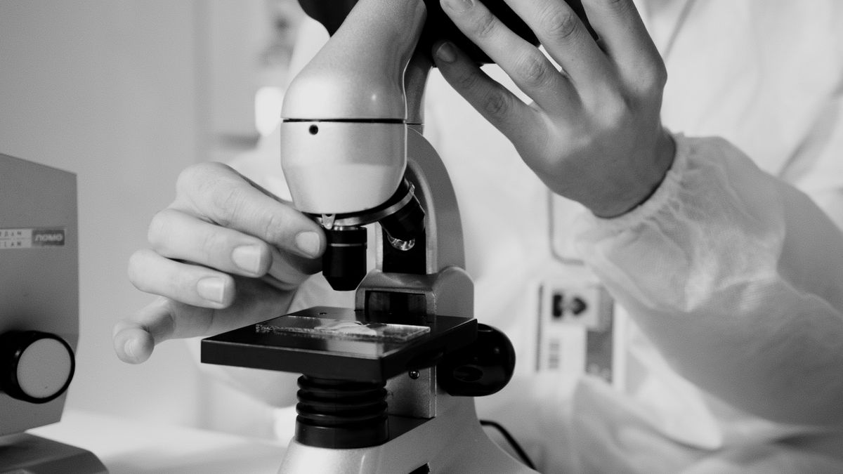
A microscope works by using two sets of lenses — the objective and the eyepiece — to magnify tiny objects that are invisible to the naked eye. Light passes through or reflects off the specimen, the objective lens creates an enlarged real image inside the microscope tube, and the eyepiece lens magnifies that image further for your eye. The total magnification is simply the objective power multiplied by the eyepiece power: a 10× eyepiece with a 40× objective gives 400× magnification.
Here is what we cover: how does a microscope work from first principles, the role of each part, how to calculate magnification, the difference between magnification and resolution, and a quick guide to using one.
How Does a Microscope Work? The Core Mechanism
Picture a slide projector. A bright light shines through a small transparent slide, a lens collects the light and projects a large image on the wall. A compound microscope does the same thing, except it uses a second lens to let you view the projected image up close.
The specimen sits on a glass slide on the stage. Light from below passes through the specimen. The objective lens — the one closest to the slide — collects this light and bends it to form a real, enlarged, inverted image inside the microscope tube. This is the first stage of magnification. The image is real, meaning it could be projected onto a screen if you removed the eyepiece.
The eyepiece (or ocular lens) then acts like a magnifying glass. It takes that real image and magnifies it further, producing a virtual image that your eye can focus on comfortably. This is the second stage of magnification. The two-stage design is what makes a compound microscope far more powerful than a simple magnifying glass.
The Main Parts of a Microscope and Their Functions
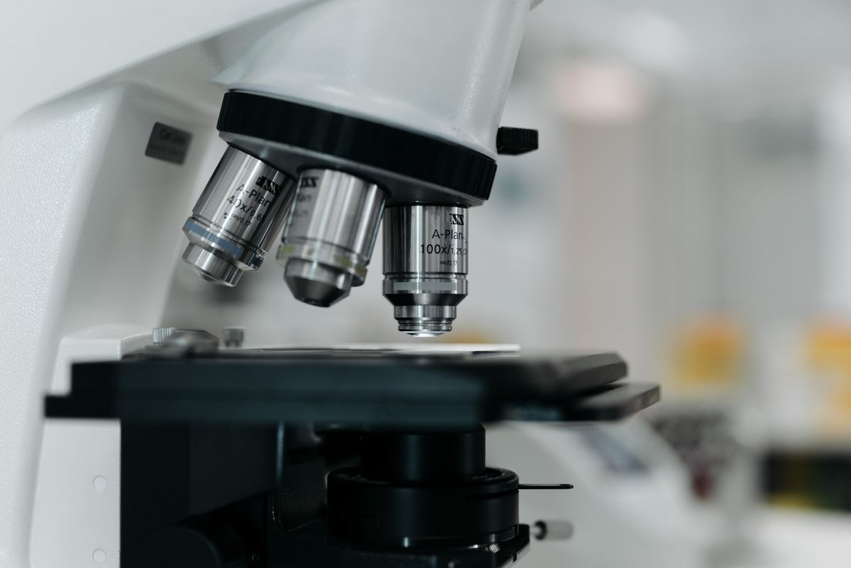
Understanding the parts is essential to understanding how does a microscope work. Each component has a specific job:
| Part | Function |
|---|---|
| Eyepiece (Ocular Lens) | The lens you look through. Magnifies the real image from the objective, typically 10× |
| Objective Lenses | The primary magnifying lenses. Usually 3-4 on a rotating nosepiece: 4×, 10×, 40×, 100× |
| Revolving Nosepiece | Holds the objectives and lets you switch between magnifications |
| Stage | The flat platform where the glass slide sits |
| Stage Clips | Hold the slide in place |
| Condenser | Focuses light from the source onto the specimen for even illumination |
| Diaphragm / Iris | Controls how much light reaches the specimen |
| Coarse Focus Knob | Moves the stage up and down for initial focusing |
| Fine Focus Knob | Makes tiny adjustments for sharp, clear focus |
| Light Source | Illuminates the specimen from below (LED or halogen) |
| Arm | The curved part you carry the microscope by |
| Base | The bottom that supports the whole instrument |
The condenser and diaphragm sit below the stage. The condenser concentrates light onto the specimen; the diaphragm adjusts brightness. Too much light washes out detail, too little leaves the image dim.
Magnification: How the Two Lenses Work Together
Magnification happens in two stages:
Stage 1 — The objective lens produces a real, inverted image that is 4×, 10×, 40×, or 100× larger than the actual specimen. This image forms inside the microscope tube at a point called the intermediate image plane.
Stage 2 — The eyepiece lens takes that intermediate image and magnifies it further, typically by 10×. The final image your eye sees is virtual — it appears to be behind the lens, similar to how a magnifying glass shows an enlarged image behind itself.
Total magnification = objective magnification × eyepiece magnification
A standard setup with a 10× eyepiece and 40× objective gives 400× total magnification. With a 100× oil-immersion objective, the same eyepiece gives 1000×.
This two-lens system is the heart of how does a microscope work. A single lens cannot reach high magnifications without severe distortion. The compound design splits the work, letting each lens do what it does best.
Resolution vs Magnification: Why Bigger Is Not Always Better
It is tempting to think higher magnification is always better. It is not. Resolution — the ability to distinguish two close points as separate — matters more.
The resolution limit of a light microscope is about 0.2 micrometres (200 nanometres). This is a hard limit set by the wavelength of visible light. Ernst Abbe discovered this in 1873: no optical microscope can resolve details smaller than roughly half the wavelength of the light it uses.
Blue light has a wavelength around 450 nm. The best resolution is about 200-250 nm. If you try to magnify beyond 1000-1500× on a standard light microscope, you get empty magnification — the image gets bigger but no new detail appears. It is like zooming in on a low-resolution photo: the pixels just get larger.
This is why electron microscopes exist. By using electrons with wavelengths thousands of times shorter than light, they achieve resolution down to 0.1 nm — about 2000 times better than any light microscope.
How to Use a Microscope in 5 Steps
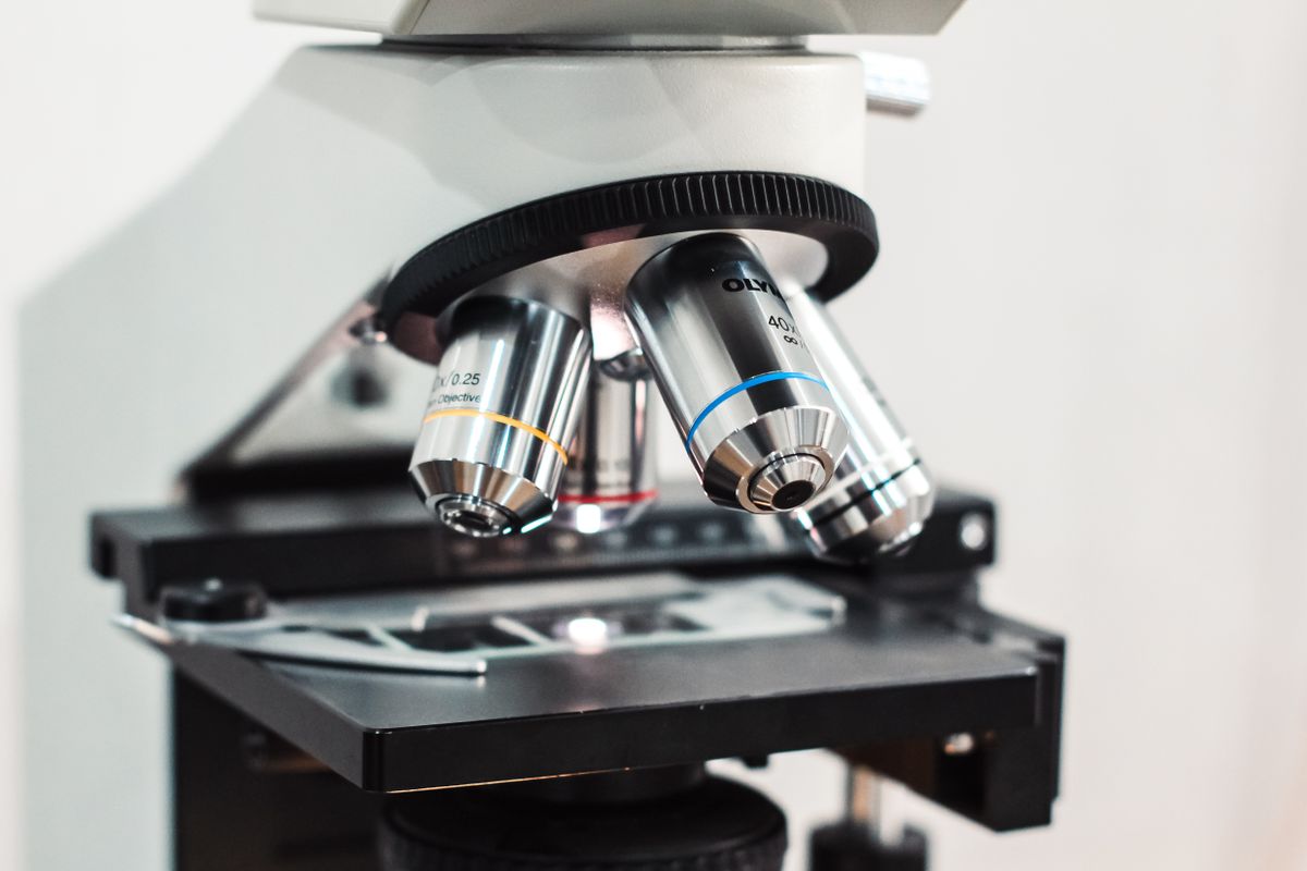
Knowing how does a microscope work in theory is one thing. Using it correctly is another. Here is a quick practical guide:
- Start low. Turn the nosepiece to the lowest-power objective (usually 4×). This gives the widest field of view and makes finding the specimen easy.
- Position the slide. Place the slide on the stage, specimen centred over the light hole. Secure it with the stage clips.
- Focus with the coarse knob. Looking from the side (not through the eyepiece), lower the objective close to the slide using the coarse focus knob. Then look through the eyepiece and turn the coarse knob to bring the image into rough focus.
- Adjust lighting. Open the diaphragm to let in more light. Adjust the condenser height for even illumination. The image should be bright but not washed out.
- Refine and increase. Use the fine focus knob for sharp detail. Switch to higher objectives as needed. Only use the fine focus knob with 40× and 100× objectives to avoid crashing the lens into the slide.
A common beginner mistake is going straight to high power. Always start at low power to locate your specimen, centre it in the field of view, then increase magnification.
Types of Light Microscopes
The standard brightfield microscope described above is the most common type, but several other light microscope techniques exist:
- Phase contrast: Enhances contrast in transparent specimens (like living cells) by converting phase differences into brightness differences.
- Dark field: Lights the specimen from the side so only scattered light enters the objective, making edges glow against a dark background.
- Fluorescence: Uses fluorescent dyes and a high-intensity light source to make specific structures glow.
- Confocal: Uses a laser and pinhole to reject out-of-focus light, producing sharp optical sections through thick specimens.
For a deeper look at how individual lenses create images, see how a lens works. The principles of refraction are what make every lens — including microscope objectives — bend light to form an image. For the fundamentals of light itself, that article covers the wave nature that limits resolution.
The Nikon MicroscopyU site offers a detailed reference on microscope optical systems covering numerical aperture, depth of field, and Köhler illumination. For the history of how the microscope evolved from a simple magnifier to a precision instrument, the Britannica microscope entry provides a thorough account.
Understanding how does a microscope work opens the door to the microscopic world. Start with the parts, understand the two-stage magnification, respect the resolution limit, and the rest follows.
Frequently Asked Questions
How does a microscope work in simple terms?
A microscope uses two lenses to make tiny objects look bigger. The objective lens (near the specimen) creates a magnified image. The eyepiece lens (near your eye) magnifies that image further. Light shines through or reflects off the specimen, travels through both lenses, and your eye sees a much larger version.
What are the 5 steps of how a microscope works?
- Light from a source illuminates the specimen on the stage. 2) Light passes through or reflects off the specimen. 3) The objective lens collects the light and forms a magnified real image inside the tube. 4) The eyepiece lens further magnifies that image. 5) Your eye sees the final enlarged image. Total magnification equals objective magnification times eyepiece magnification.
What is the difference between magnification and resolution?
Magnification is how much larger an object appears. Resolution is how clearly you can see fine detail — the ability to distinguish two close points as separate. A microscope can magnify 1000×, but if the resolution is poor, the image is just a big blur. Resolution is limited by the wavelength of light to about 0.2 micrometres for optical microscopes.
What are the main parts of a microscope?
The main parts are: eyepiece (ocular lens), objective lenses (usually 3-4 on a revolving nosepiece), stage (holds the slide), condenser (focuses light onto the specimen), diaphragm (controls light amount), coarse and fine focus knobs, arm, base, and light source.
How do you calculate total magnification?
Multiply the eyepiece magnification by the objective magnification. If the eyepiece is 10× and the objective is 40×, the total magnification is 10 × 40 = 400×. Most compound microscopes have eyepieces of 10× and objectives of 4×, 10×, 40×, and 100×, giving total magnifications from 40× to 1000×.
What is a compound microscope?
A compound microscope uses two or more lenses to magnify an object. The objective lens produces a real, inverted, magnified image inside the tube. The eyepiece lens then magnifies this image further. Most modern microscopes are compound microscopes. Simple microscopes (like a magnifying glass) use only one lens.
How is a light microscope different from an electron microscope?
A light microscope uses visible light and glass lenses to form an image, with a maximum resolution of about 0.2 micrometres and magnification up to 1000-1500×. An electron microscope uses a beam of electrons and electromagnetic lenses, achieving resolution down to 0.1 nanometres and magnification over 1,000,000×. However, electron microscopes require vacuum, complex sample preparation, and cannot image living specimens.

About Umar Farooq
Umar Farooq writes in-depth guides on the physics of light and optics — from reflection, refraction, and lenses to diffraction, lasers, and fiber optics, explained from first principles.
Read more about Umar Farooq →